
Fluid Overload After Liposuction: Causes, Prevention, and When to Seek Help

Key Takeaways
- Fluid overload after liposuction occurs because tumescent fluid and tissue trauma exceed local lymphatic drainage. Keep an eye on treated regions for lingering swelling or soft, movable fluid pockets.
- Differentiate normal postoperative swelling from abnormal accumulation by monitoring persistence, severity, and indications such as serous fluid leakage, increasing pain, or recurrent nodules.
- Determine personal risk through medical history, procedure scale, and anesthesia type, and prudently modify the plan when heart, kidney, or liver disease, prior seromas, or large-volume or revision procedures are present.
- Prevent overload through careful preoperative screening, precise intraoperative fluid calculation and monitoring, and postoperative measures such as compression garments, early lymphatic drainage, and scheduled follow-up.
- Surgeons should determine maximum tumescent volumes safe for their liposuction technique, chart fluid intake and output, educate staff to recognize warning signs, and provide patients with explicit recovery guidance and emergency contacts.
- Urgent care for serious or worsening symptoms such as shortness of breath, chest pain, high fever, rapidly expanding fluid collections, confusion, or syncope.
Fluid overload after liposuction recognition and prevention refers to identifying excess fluid buildup and taking steps to stop it. Symptoms are swelling, shortness of breath, and weight gain within days after surgery.
Risk increases with large-volume procedures, intravenous fluids, and pre-existing heart or kidney problems. Close vital sign monitoring, judicious fluid limits, and clear post-operative instructions decrease risk.
The body details evaluation, thresholds, and hands-on preventative steps.
Understanding The Cause
Fluid overload after liposuction is due to a combination of local changes in the tissue and systemic fluid shifts. Surplus tumescent fluid, tissue trauma, disrupted lymph flow and supplemental IV fluids all come together to raise interstitial fluid. The lymphatic system washes away excess fluid under normal conditions, but when it is overwhelmed or injured, fluid pools and can form seromas or chronic edema.
Tumescent Fluid
Tumescent fluid is injected to numb the area, minimize bleeding, and provide a plane in which to remove fat. Correct volumes accelerate the process and minimize discomfort. Excess fluid increases interstitial pressure and can become encased in fat pockets.
Improper dosing or not considering patient size results in fluid absorption beyond lymphatic and renal clearance, generating fluid overload. Tumescent technique is completely different from wet, superwet, and large volume by volume and anticipated absorption. Superwet and large-volume liposuction use significantly more fluid and thus have a higher risk of retained tumescent solution if not balanced with cautious monitoring.
Tumescent fluid can lead to prolonged swelling, seroma pockets, and delayed healing, which may require drainage or compression to resolve. Complications related to retained tumescent fluid can be visible swelling, localized pain, seroma, and an increased risk of infection if the fluid becomes secondarily infected.
A patient with extensive abdomen and flank liposuction who received large tumescent volumes may present with fluctuating swell and pockets that require ultrasound-guided aspiration.
Body’s Response
Surgical trauma sets off an inflammatory cascade. After injury, capillary permeability is increased by inflammatory mediators, physical injury, ischemia or drugs, enabling plasma proteins and fluid to leak into interstitial spaces. This leak causes swelling and can inundate local lymphatic channels.
Lymphatic drainage is precarious. Small vessels can be cut, crushed, or thrombosed during cannula passage. When lymph flow is low, fluid washes slowly. Secondary lymphedema can arise when blocked channels are unable to return fluid. This increases risks of subcutaneous compartment buildup and seroma.
Capillary endothelium to stress and crystalloids. Iatrogenic hemodilution from IV crystalloids increases capillary leak and decreases plasma colloid osmotic pressure, enhancing fluid shift into tissues. Surgical patients who receive extra IV saline or large crystalloid boluses may have worse edema than those managed with a judicious fluid approach.
Surgical Factors
Amount and depth of liposuction are important. Excessive fat extraction or deep lipectomy leaves more dead space and trauma, which encourages more fluid accumulation. Revision procedures and multiple treated areas compound the risk because repeat trauma further damages lymphatics.
Good surgical planning limits fluid overload. Choose an appropriate technique, limit tumescent and IV crystalloid volumes, stage procedures if large areas are needed, and use compression postoperatively.
Restricting intraoperative fluid ratios and unnecessary crystalloids decreases systemic hemodilution and capillary leak. With lymphatic obstruction or patient comorbidity, anticipate elevated edema danger and strategize.
Recognizing The Signs
Fluid overload after liposuction can be anywhere from mild, anticipated swelling to a significant complication requiring immediate treatment. The following subsections identify the outward signs, how to use rudimentary vital signs to detect trouble, the internal imbalances to look for, and how to differentiate normal postoperative changes from abnormal fluid retention.
1. Physical Symptoms
External symptoms are localized lumps, bumps, and constant swelling in the treated areas. Small, soft pockets under your skin that feel squishy and shift a bit when you press them are usually seromas and can make an appearance within the initial weeks post-surgery, typically around day 7 to 10 or when drains are taken out.
Draining clear fluid from incision sites or soft compressible bulges adjacent to the liposuction sites are red flags and need to be evaluated. Seromas can occur weeks post-op. If new swelling develops a few weeks after surgery or a previous swelling worsens, think seroma.
Pain that remains intense or becomes localized on or after two weeks after surgery is another warning sign that a fluid pocket may have formed instead of normal healing soreness. Persistent or worsening swelling is NOT your typical post-op puffiness. It lasts longer and has a different feel.
Normal swelling reaches its peak in the first week and then gradually subsides. If the region remains notably edematous, develops fluctuance, or has purulent drainage, aspiration and clinical evaluation are usually required to diagnose and manage the collection.
2. Vital Signs
Be alert for signs of intravascular overload. Monitor blood pressure and heart rate. High blood pressure with a bounding pulse can be indicative of excess circulating fluid. If he is breathing fast or is short of breath, that could indicate fluid has shifted into the lungs, which is pulmonary edema, and requires immediate treatment.
Monitor for signs of infection surrounding seromas, as infected collections of fluid alter management. Create a daily log of vitals during the early recovery period. Record blood pressure, pulse, respiratory rate, and temperature once or twice daily to spot trends early.
3. Internal Imbalances
Too much can dilute sodium and potassium, making you weak, confused, or crampy. Urinary output changes, either significantly decreased or inappropriately dilute, indicate fluid and electrolyte shifts and must be communicated.
Fluid overload may cause acidosis or pulmonary symptoms, such as cough, difficulty breathing, or decreased exercise tolerance. Among severe fluid shifts, fluid overload and fluid depletion are risk factors for fat embolism and blood clots after liposuction, which are rare but serious and require immediate medical treatment.
4. Normal vs. Abnormal
We anticipate swelling to be at its highest in week one and to steadily improve. Abnormal buildup is lingering, recurring, or ascending after an initial descent. Mild seromas are small, transient and may resolve.
Large or recurrent seromas require aspiration, imaging or drainage. When to seek treatment: Duration, pain worsening after 2 weeks, leaking clear fluid and soft, moveable lumps require intervention. Consult concerning milestones in normal healing versus warning signs in persistence to determine when to seek care.
Aspiration is the optimal diagnostic test for suspected seroma.
Assessing Your Risk
Evaluating risk for fluid overload post-liposuction involves thoughtful consideration of patient parameters, surgical strategy, and anesthetic decisions. This establishes the foundation for fluid management, monitoring, and post-op care decisions.
Medical History
Chronic illnesses that impair fluid balance raise risk. Heart failure, chronic kidney disease, and advanced liver disease reduce the body’s ability to handle extra fluid and can lead to pulmonary edema or worsened edema postoperatively.
Prior surgeries are a concern, too. Patients with previous seroma formation, multiple liposuction procedures, or wide scarring have disrupted lymphatic drainage and increased fluid accumulation.
Varicose veins and lymphatic disorders warrant attention as inadequate venous or lymphatic flow prolongs local swelling and increases the risk of complications. Document all medications.
Diuretics, ACE inhibitors, NSAIDs, anticoagulants, and steroids change fluid status or bleeding risk and should be recorded and discussed before surgery. They need a clear, written medication list because it helps the team plan intraoperative fluids and postoperative monitoring.
Procedure Scale
Greater-volume liposuction and multiple areas treated add to tissue trauma and inflammatory reaction, which increases local and systemic fluid shifts. Primary versus revision cases differ.
Revisions often require more dissection and scar release, increasing bleeding and fluid sequestration. More aggressive fat removal techniques, such as power-assisted or ultrasound-assisted methods, increase the risk of contour irregularities and higher fluid requirements due to additional tissue trauma.
The correlation between procedure size and fluid management needs follows a dose-response trend. Bigger procedures predict larger fluid shifts and higher IV fluid requirements. This is probabilistic rather than absolute.
Tumescent liposuction under 4 L of supranatant fat and under 3% of body weight generally doesn’t require IV fluids. Veteran surgeons include anticipated blood loss, contour risk, and likely fluid shifts in the equation.
| Procedure scale | Typical concerns | Expected fluid management needs |
|---|---|---|
| Small, <4 L tumescent | Low blood loss, minimal trauma | Often no IV supplementation |
| Moderate, multi-area | Moderate trauma, possible seroma | Monitor; selective IV support |
| Large or revision | High tissue trauma, higher bleeding | Proactive IV fluids, close monitoring |
Anesthesia Type
Local, tumescent, and general anesthesia all alter the way fluids are administered and how symptoms manifest. Tumescent and superwet perform local fluid administration; they require careful accounting to prevent overuse.
Superwet and tumescent need to be dosed by weight and area and can lead to systemic overload if miscalculated. General anesthesia masks early indicators such as breathlessness; therefore, clinicians must depend on observation instead of patient feedback.
Adjust intraoperative supplementation by anesthesia: minimal IV for pure tumescent small cases, measured IV for larger or mixed techniques, and vigilant fluid control with general anesthesia. Play it safe around 21 mm Hg when budgeting to cut down pulmonary edema danger.
Experienced surgeons are better at recognizing and reacting to these problems as they develop.
Proactive Prevention
It’s important to note that preventing fluid overload starts with a plan before, during, and after liposuction. To restrict fluid influx, track equilibrium in real time, and facilitate natural post-op fluid elimination. The steps below are numbered to reflect practical actions clinicians and patients can take.
- Preoperative assessment and optimization.
- Precise intraoperative fluid strategy and technique.
- Structured postoperative care, monitoring, and education.
- Early interventions for swelling, seroma, or infection.
- Active follow up to prevent fibrosis and chronic swelling.
Preoperative Screening
Need to measure baseline hydration status and electrolyte levels via blood tests and clinical exam. Check serum sodium, potassium, creatinine, and hemoglobin to flag hemodilution risk or renal issues that affect fluid handling.
Mandate evaluation for conditions that alter fluid balance: heart failure, liver disease, renal insufficiency, thyroid disease, uncontrolled diabetes, and hypoalbuminemia. NSAIDS, steroids, and diuretics change fluid distribution.
Checklist: medical history, vitals, BMI, lab panel, cardiac and renal consults if indicated, medication review, and informed consent about fluid risks. Employ a preoperative checklist to get everything signed off and talked through.
Provide patient guidance: avoid NSAIDs preoperatively as advised, maintain normal hydration but not intentional overdrinking, and limit strenuous activity in the two weeks before surgery to reduce inflammatory baseline.
Intraoperative Care
Nits: 1) Calculate and cap tumescent fluid volumes exactly according to body area and aspirate estimate. Utilize the smallest effective cannula, frequently under 3 mm, to minimize tissue trauma and fluid sequestration.
Monitor fluid input and output in real time: record tumescent inflow, IV fluids, and measured aspirate. Monitor urine output, heart rate, and blood pressure to catch early hemodilution.
Use technology, such as ultrasound-assisted or power-assisted liposuction, to minimize manual trauma and reduce fluid requirements. Over-hydrate with IV fluids only if hypotensive or if there is documented blood loss.
Antibiotic prophylaxis may be warranted. Agents like ciprofloxacin are employed in certain regimens to minimize infection risk that can exacerbate edema.
Postoperative Management
Use compression garments routinely and consider bimodal compression: firmer wrap for the first 24 to 72 hours, then graduated garment to control swelling. Promote gentle movement for several weeks.
Light walking aids circulation, but don’t do anything too vigorous to increase bleeding or inflammation. Initiate lymphatic drainage massage as soon as possible, sometimes within 24 to 72 hours, to increase fluid mobility and decrease seroma.
Watch for seroma, infection, or persistent swelling at follow-ups and treat promptly. Teach patients about healthy hydration, not overhydration, and unambiguous wound care, pain management, and red flags to communicate.
Start fibrosis prevention treatments within 24 hours and continue for weeks to months as needed.
The Surgeon’s Role
Surgeons should assume first accountability for liposuction fluid management. That responsibility mixes artistry in form shaping with meticulous, science-based perioperative care. A surgeon expert in liposuction and body contouring straddles art, smooth contours and balanced proportions, with clear medical judgment regarding fluids, risks and early signs of complication.
Fluid Calculation
Proper fluid math starts prior to the initial incision. Calculate the maximum safe tumescent volume based on patient weight and the number of areas treated. For example, use conservative limits of tumescent per kilo with a stepwise increase for every additional treatment area, adjusted for comorbidities.
Adjust IV crystalloid rates and tumescent infiltration for each zone. Larger areas or combined procedures require lower per-area infiltration to avoid cumulative overload. Record total intraoperative and postop I/O. Note tumescent volume infiltrated, aspirate volume, estimated blood loss, and all IV fluids.
Monitor urine output hourly during long cases. Use a simple table to display recommended maximum tumescent volumes by weight and procedure scope:
| Patient weight (kg) | Single small area (e.g., abdomen only) | Multiple areas (e.g., abdomen + flanks) | Extensive body work (abdomen + flanks + thighs) |
|---|---|---|---|
| <60 | 20–30 mL/kg | 15–25 mL/kg | 10–20 mL/kg |
| 60–80 | 15–25 mL/kg | 12–20 mL/kg | 8–16 mL/kg |
| >80 | 12–20 mL/kg | 10–18 mL/kg | 6–14 mL/kg |
Adjust these ranges for age, cardiac or renal disease, and combined procedures such as BBL.
Patient Monitoring
Ongoing evaluation is required from recovery into early outpatient follow-up. Observe for advancing swelling, tense seromas, rising respiratory rate, hypoxia, or new crackles which could indicate pulmonary edema from excessive IV fluids.
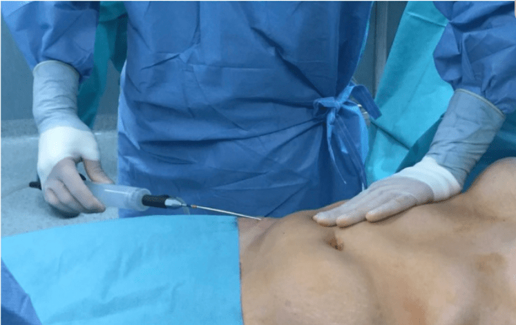
Follow-ups should occur at 24 to 48 hours, one week, two weeks, and additional visits based on the findings. Train staff to spot subtle cues: disproportionate unilateral swelling, rapid weight gain, or decreasing urine output.
Record each evaluation, treatment, and patient statement on the chart for trend analysis and legal transparency.
Transparent Communication
Explain risks and prevention steps plainly: why tumescent limits exist, signs of seroma versus normal postoperative swelling, and when to seek help. Provide written directions of what the red flags are: shortness of breath, chest tightness, sudden large-area swelling, and low urine output.
Include clear phone numbers for concerns that are urgent. Support truthful, early patient feedback throughout post-operative recovery and provide a low threshold for early clinic review or scanning.
It’s the part of communication that we frame as partnership, boosting follow-up adherence and catching fluid overload early.
When To Seek Help
Fluid accumulation post-liposuction can begin insidiously or present acutely within days. Too little detail can lead to missing the signs and symptoms that should trigger a visit to the doctor. Fluid pockets, or seromas, generally develop approximately five to ten days post-surgery. Early follow-up and consistent wear of a compression garment minimize risk, but issues can still arise and require prompt care.
Seek emergency care for serious symptoms. Prompt evaluation is required for any new or worsening shortness of breath, chest pain, sudden rapid swelling, severe pain, fainting, or confusion. These signs may point to either fluid overload, pulmonary issues, or other emergencies requiring urgent treatment. Don’t wait for your appointment. Call 911 or head to the closest hospital.
Inform your surgeon of any recurrent or quickly expanding fluid pockets or seromas. If you observe a mushy, fluctuating swelling that increases in size over the days, or a collection that was small and is now becoming larger, get in touch with your surgical team. Seromas usually manifest themselves five to ten days post-operation but may be delayed. Your surgeon can typically detect an early seroma on exam and determine whether conservative care, needle aspiration, or a small drain is required.
There’s care for fluid leakage or draining wounds. Seromas will occasionally seep clear or slightly yellow fluid through incision sites. If you notice persistent drainage, wet dressings or a dressing that soaks through time and again, seek medical attention. Persistent leakage can increase infection risk and may necessitate aspiration, wound care, or temporary drainage.
Be on the lookout for infection at incision sites. If the redness spreads, warmth intensifies, there is pus, fever, or increasing pain, get it checked. Infections can masquerade as seromas or come on the heels of them. Early antibiotic treatment and potential drainage prevent complications.
Follow directions on compression garments and drains. Compression garments should be worn day and night in the early recovery phase. Taking them on and off frequently or not wearing them increases your risk of seroma formation. Drains, if utilized, are typically removed within a few days to a week. If you note increasing drainage, new swelling, or changes in the character of the fluid after drain removal, reach out to your surgeon.
Go to follow-ups and report symptoms that persist. Some swelling and seromas dissipate over weeks to months. Continued or increasing swelling, pain, or drainage beyond this should prompt re-evaluation. Your surgeon can provide interventions to accelerate recovery and avoid chronic problems.
Conclusion
Fluid overload after liposuction can make an appearance quickly. Be on the lookout for swelling that continues to increase, tight skin, shortness of breath, or rapid pulse. Monitor drain output, weight, and your symptoms. Low-risk steps help cut the chance: limit IV fluids, use drains when needed, and follow post-op rest and salt advice. Surgeons have an important role through preoperative fluid planning and close postoperative patient monitoring. Receive care immediately for any sudden difficulty in breathing or swelling that fails to subside.
A good hand, regular monitoring and open conversations with your medical team do matter. If concerned about fluid overload, call your surgeon or go to urgent care now!
Frequently Asked Questions
What is fluid overload after liposuction?
Fluid overload is extra fluid in the body post-surgery. It can be the consequence of massive intra-operative volumes or inadequate aspiration and it impacts respiratory, circulatory, and wound healing processes.
What are the earliest signs to watch for?
Early symptoms involve sudden swelling, shortness of breath, rapid weight gain, and a sensation of tightness in the chest or abdomen. Report these immediately to your surgeon or emergency care.
Who is at higher risk for fluid overload?
Higher risk includes patients with heart, kidney, or liver disease, older adults, large-volume liposuction, and patients receiving large intraoperative fluid volumes. Talk about risks at your pre-op.
How can I reduce my risk before surgery?
Minimize risk by providing your complete medical history, discontinuing medications as directed, bringing any chronic conditions under control, and selecting a board-certified surgeon with extensive experience managing fluids and using tumescent techniques.
What intraoperative measures prevent fluid overload?
Surgeons avoid overload with meticulous tumescent technique, restrictions on total fluid volumes, monitoring of vital signs, and drain placement when appropriate. Veteran crews monitor fluid in and out.
What should I do if I suspect fluid overload after discharge?
Get immediate medical care. Contact your surgeon or your emergency department if you experience breathing difficulty, chest pain, rapid swelling, or fainting. Early treatment leads to better outcomes.
Can fluid overload be treated and fully resolved?
Yes. Treatment can involve diuretics, oxygen, IV medicines, and hospital observation. With early recognition, prompt treatment generally results in full recovery.
 Directions
 Call Us
 Email Us