
Protectin and Tissue Protection After Liposuction: Role, Risks, and Recovery Best Practices
Key Takeaways
- Liposuction induces controlled subcutaneous trauma. Therefore, emphasize gentle techniques, careful cannula manipulation, and appropriate fat extraction amounts to minimize tissue damage and optimize recovery.
- Protectin is a lipid mediator that resolves inflammation, protects cells from oxidative stress, and promotes tissue regeneration. This potentially enhances fat graft survival and minimizes scarring.
- Combine these inflammation-controlling, cell-protective strategies with scar prevention in postoperative protocols using inflammation modulating therapies, careful wound care, and proven scar management techniques.
- Track patients postoperatively with objective tissue health metrics and investigate protectin as a biomarker to inform personalized postoperative management and early intervention.
- Pair protectin-based therapies with regenerative approaches, lymphatic drainage, pain management, and lifestyle measures such as compression, gentle exercise, and smoking cessation.
- Remember to keep whole-minded, combining molecular interventions with surgical excellence, atraumatic instrumentation, port protection and multidisciplinary post-operative care to maximize results and patient satisfaction.
Protectin and tissue protection after liposuction science refers to how protectin molecules support healing and reduce inflammation after fat removal. Protectins are derived from omega-3 fats and assist in clearing cellular debris, reducing inflammation, and directing tissue protection.
Research demonstrates they function at micro doses and complement light massage, compression, and nutrition. The bulk reviews mechanisms, clinical science, dosing considerations, and practical advice to support recovery.
Liposuction Trauma
Liposuction creates measured trauma in subcutaneous tissue, fat cells, small blood vessels, lymphatics, and extracellular matrix. The surgery specifically tears up the fat layer to extract the fat cells, but that tearing also induces bleeding, inflammation, and fluid shifts.
The first week is the worst with swelling and bruising. Inflammatory cells are present within days and actual remodeling of the tissue can persist for months. Early changes drive contour and sensation outcomes, which is why technique and post-op care mold both short- and long-term results.
The Procedure
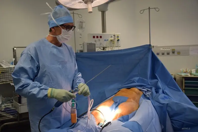
Liposuction generally commences with anesthesia and small incisions. Surgeons infiltrate tumescent solution to minimize bleeding, which facilitates fat suction, then insert a cannula that they agitate to fracture fat, which is suctioned via tubing, while port protectors and incision mapping reduce skin trauma.
Tumescent liposuction uses huge volumes of dilute local anesthetic and epinephrine to minimize blood loss. Assisted techniques, including ultrasound-assisted, power-assisted, or laser-assisted, apply energy or mechanized motion to break up fat prior to suction.
Precise fat removal volumes matter: over-resection causes contour deformity and under-resection leaves unevenness. When fat grafting takes place thereafter, gentle handling, such as Coleman-style harvesting, centrifugation, and slow injection, strives to maintain fat cell viability and reduce secondary tissue trauma.
Tissue Response
Right after liposuction, a flood of fluid and inflammatory mediators leads to swelling and bruising, which peak in week 1. Hemostasis begins immediately, inflammation persists for days, succeeded by proliferation with fibroblast and capillary ingrowth.
Remodeling of collagen and fat architecture may extend over several months. Scar tissue and collagen deposition are a component of repair. If unbridled fibrosis and adhesions can form and tether skin to deeper tissues, lymphatic channels are disrupted, which can generate seroma or persistent edema.
Lymphatic drainage massage, proper wound care, and wearing compression garments most of the day for 6 weeks or so reduce fluid retention and assist with sculpting as tissues reconfigure.
Common Complications
- Swelling and bruising (most pronounced in first week)
- Seroma (fluid collection)
- Infection and delayed wound healing
- Contour irregularities and surface rippling
- Fat necrosis and calcification
- Adhesions and fibrosis causing restricted mobility
- Fat embolism and visceral injury (rare but serious)
Aggressive suction, excessive cannula motion, or poor technique increases the risk of complications including visceral perforation and fat embolism syndrome. Surface irregularities usually originate from uneven fat removal or unrecognized adhesions.
Early detection and care, such as drainage of seromas, antibiotics for infection, and targeted massage, reduce long-term problems. Compression, staged procedures, and gentle intraoperative handling all reduce complication rates and enhance the final contour.
Protectin’s Role
Protectin is one of the lipid mediators produced during inflammation resolution with obvious anti-inflammatory and tissue-protective activities that could be important to liposuction recovery. It intervenes at several points in the healing cascade to reduce damage, maintain cell viability, and direct rational repair. Among these, mechanisms are organized into the critical functional domains that impact surgical outcomes and patient care.
1. Inflammation Control
Protectin downregulates pro-inflammatory cytokines like IL-1β, TNF-α, and IL-6 and limits early neutrophil influx into the surgical site. This transition diminishes acute inflammation’s magnitude and duration after fat excision. Keeping inflammation in check reduces swelling and pain and decreases the likelihood of hypertrophic scarring.
With inflammation under control, fewer adipocytes get exposed to damaging enzymes and free radicals that can degrade fat cells and subcutaneous structures during those key settling weeks and months. Protectin’s role in integrating inflammation-modulating strategies — pharmacologic agents, timing of interventions, and compressive therapy — supports a calmer healing environment and complements the role of compression garments that provide external pressure and reduce edema.
2. Cell Protection
Protectin protects fat cells, fibroblasts, and endothelial cells from oxidative stress and apoptosis by inducing pro-survival signaling and antioxidant responses. This cellular protection enhances fat graft retention and preserves microvascular integrity, both of which increase tissue quality and long-term contour stability.
Less cell death decreases the amount of fat necrosis and oil cysts and reduces aberrant healing responses that cause irregularities. Tracking tissue health via clinical examination, ultrasound when indicated, and patient-reported outcomes helps evaluate the effectiveness of these cell-protective interventions in addition to conventional post-op care such as compression garments for several weeks.
3. Tissue Regeneration
Protectin drives regeneration of damaged skin, fascia, and subcutaneous fat by supporting debris-clearing and growth factor-releasing macrophage phenotypes. Rapid, coordinated regeneration facilitates even tissue re-adhesion and efficient skin retraction, which is key to smooth contouring and minimal downtime.
Regenerative adjuncts, including PRP, controlled physical therapy, and compression, can cooperate with protectin pathways to promote endogenous repair. The first weeks are where we see the most change. It takes months before the final shape starts becoming visible as the swelling goes down and tissues begin to settle.
4. Scar Prevention
Protectin disrupts excess collagen deposition and myofibroblast over-activity, reducing the risk of hypertrophic scars and deep adhesions. Less abnormal scar tissue means better surface appearance and better subcutaneous glide, less tethering, and firmness.
For best results, combine protectin-focused approaches with established scar management: massage, silicone sheeting, and compression garments to limit tissue movement and guide collagen remodeling.
5. Pain Reduction
Protectin has analgesic properties that reduce postoperative soreness by dampening nociceptive signaling linked to inflammation. Less pain enables earlier mobility and better patient compliance with lymphatic drainage massage and gentle activity, which in turn supports recovery.
Pain control paired with compression garments helps patients manage limited mobility and fatigue in the initial days, making routine care easier and improving overall convalescence.
Clinical Significance
Protectin, an endogenous lipid mediator associated with resolution of inflammation, is increasingly relevant for tissue protection post-liposuction. There’s clinical significance with evidence of benefit in reducing inflammation, improving fat graft survival, limiting contraction and scar formation.
With the proliferation of energy-based liposuction methods, which expose patients to increased risks of thermal injury and port-site scarring, adjunctive strategies that harness protectin’s actions are clinically significant.
Patient Outcomes
| Outcome domain | Traditional recovery | Recovery with protectin-based care |
|---|---|---|
| Overall complication rate | 33.3% (no port-site protection in one review) | Reduced to |
| 6.67% with port-site protection reported in retrospective review |
| Portsite complications | Thermal burns, wound dehiscence, and infection reported. Less common, but 6.67% still experienced in the shielded cohort |
| Revision surgery rate | As many as 13.3% had multiple revisions in a single study. Fewer revisions were anecdotally seen where protection/protectors were utilized. |
| Patient satisfaction | 76 to 86 percent based on pain, weight gain, complications | There is a tendency toward greater satisfaction when complications are lower |
When compared to standard recovery, protectin-based protocols demonstrate more defined early tissue preservation and reduced salvage measures in retrospective and preclinical models.
Protectin seems to aid fat graft take by preventing local inflammatory cell influx and limiting necrosis, thereby decreasing subsequent contour revision requirements.
Patient-reported satisfaction continues to be key. Objectively quantified measures such as scar grade, tissue elasticity, and volume retention of grafts should be complemented with validated satisfaction surveys to capture outcomes that are both functional and aesthetic.
Practical steps involve standardized wound photography, scar scales, and patient-reported outcome measures at regular time points.
| Outcome Measure | Technique A | Technique B | Technique C |
|---|---|---|---|
| Complication Rate (%) | |||
| Revision Rate (%) | |||
| Pain Score (0-10) | |||
| Graft Survival Rate (%) |
Biomarker Potential
Protectin concentration in tissue or plasma may be promising as a predictor of healing capacity and complication risk post-liposuction. Early low levels might flag patients who will develop a prolonged inflammatory response and experience higher rates of wound breakdown or poor graft survival.
Using protectin as a guide could let clinicians tailor postoperative care. Patients with low levels receive enhanced anti-inflammatory regimens, closer follow-up, or protective measures such as port-site protectors.
Non-invasive monitoring through blood tests or by assaying wound exudate is possible and would fit into routine follow-up without major burden.
Incorporating biomarker assessment into decision-making supports personalized care and could reduce overall costs by targeting interventions. The minimal cost of some protectors, around $1.87 each, contrasts with the higher expense of multiple revisions, suggesting a favorable cost benefit if outcomes improve.
Future Therapies
Future therapies will capitalize on what we’ve learned so far that the tissue recovery process after liposuction is a stepwise, multi-stage phenomenon. A brief overview helps set context: gentle fat harvest and handling reduce cell loss and trauma, early lymphatic support limits edema, and true tissue remodeling can take months. Therefore, new treatments must fit into a structured, staged protocol to be effective.
Newer treatment options that target healing of the tissue and prevention of complications include several innovative approaches.
- Localized protectin-based formulations: Topical or injectable protectin derivatives applied at the time of surgery or early post-op to reduce inflammation and protect cell membranes. These seek to reduce acute tissue damage and reduce the risk of fat necrosis. Example: A protectin gel placed in the lipoaspirate pocket to limit oxidative damage and modulate early immune response.
- Combined fat-plus-cell therapy: Loading processed fat with concentrated adipose-derived stem cell (ADSC) pellets prepared within roughly 80 minutes and mixed with fat before reinjection. Very similar to Coleman-style low-pressure harvest and gentle processing to preserve cell viability, then supplements ADSCs to support graft take and early healing.
- Growth-factor scaffolds with protectin: Biodegradable meshes or hydrogels that release growth factors and protectins over days to weeks, supporting angiogenesis and gradual tissue remodeling. Example: A collagen hydrogel that delivers VEGF and protectin to speed blood vessel ingrowth into grafts.
- Lymphatic-support bundles: Protocols that combine lymphatic drainage massage, low-pressure suction techniques, and pharmacologic lymphotrophics to reduce seroma and fibrosis. These begin immediately post-op and persist through early remodeling.
- Point-of-care cellular concentrates: Rapid bedside systems that isolate stromal vascular fraction or ADSC pellets, allowing surgeons to tailor the cell dose to patient needs in the same procedure.
- Imaging-guided delivery and monitoring: Ultrasound or near-infrared tools used intra-op to place grafts and post-op to track vascularization and detect early complications for timely intervention.
More predictable benefits will come from combining protectin with ADSCs and defined growth factors. Protectin can tone down inflammation and guard cells against oxidative stress, making for a more hospitable niche for stem cells to engraft.
ADSCs introduce pro-regenerative signals that enhance capillary growth and matrix remodeling. Together, they may optimize fat graft survival, minimize fibrosis, and accelerate restoration of tissue biomechanics. A practical example is a Coleman-style fat graft mixed with an 80-minute processed ADSC pellet and a slow-release growth factor hydrogel that contains protectin.
Surgeons should optimize low-pressure lipoaspiration, meticulous fat handling, and 3D reinjection to maintain cell viability. Post-op care must be protocolized. Early lymphatic massage, tailored compression, and staged cellular or biologic boosts are timed to the patient’s healing phase.
A Holistic View
A holistic view considers liposuction recovery an active, phased process with defined objectives at each phase. It casts healing as collaborative labor among molecular implements, the surgeon’s hand, and patient habits. This minimizes scarring, safeguards the surgical result, and provides patients with peace of mind in the weeks and months following the procedure.
Beyond Molecules
Surgical craft and soft tissue management established the platform for any molecular intervention to operate. A surgeon’s cannula size, trajectory, and suction parameters modulate local trauma and inflammatory burden. Operative environment matters: temperature control, careful fluid management, and strict hemostasis limit bleeding and hematoma risk.
Molecular agents such as protectins may be able to modulate inflammation, but they do not supplant core surgical principles. Absent atraumatic technique, pharmacology provides limited advantage. Practical device choices cut tissue stress. Silicone skin protectors at ports reduce shear on the dermis. Port protection devices prevent skin pinching.
Blunt, atraumatic instruments minimize sharp disruption. Things surgeons can do are preplan access points to avoid dense scar, use planned layered aspiration, and minimize repetitive passes in the same plane. Identify predictive biomarkers of future disease activity and disability.
Patient Factors
Create a checklist before any procedure:
- Age, BMI, and fat distribution pattern.
- Smoking status and comorbidities (diabetes, bleeding disorders).
- Medications and supplements that affect healing.
- Prior surgeries and scar patterns.
- Lifestyle factors include activity level, support at home, and ability to follow aftercare.
Customize technique and graft to every profile. Higher BMI might require staged procedures. Thin patients necessitate conservative aspiration to prevent contour deformity. Discuss expected healing: the first days bring a normal inflammatory phase.
Weeks three to five often show the first meaningful contour improvement as swelling falls. Build a three-stage regenerative plan: immediate inflammation control, early remodeling during weeks three to eight, and long-term tissue remodeling lasting months to six months and beyond.
Detailed preoperative evaluation and straightforward patient instruction minimize unanticipated occurrences. Leverage written plans that include risks, expected timelines, and when to seek care. Rerun the checklist at every follow-up to identify deviations as early as possible.
Lifestyle Impact
- Wear compression garments as advised.
- Attend scheduled follow-up visits.
- Stop smoking and using nicotine several weeks before and after surgery.
- Begin light walking within 24 to 48 hours and advance to light exercise at 2 weeks.
- Think about lymphatic drainage massage sessions beginning in week one or week two.
- Get lots of water and fuel your repair with a protein-heavy, nutrient-dense diet.
- Avoid NSAIDs when directed if bleeding risk is a concern.
Provide a simple patient guide with visual timelines: days 0 to 7 for inflammatory care, weeks 3 to 5 for visible contour change, and months 3 to 6 for ongoing remodeling. This prepares patients to anticipate a more gradual improvement and realize that complete resolution may spread over multiple months.
Enhancing Recovery
Postoperative care after liposuction determines the pace and excellence of recovery. These focused steps, timely checks, and coordinated care decrease the odds of complications and help your body adapt to its new shape. Recovery follows a predictable arc: an initial inflammatory phase in the first days, tissue repair and reorganization over the following weeks, and gradual contour refinement that can take months.
The best ways to speed healing and minimize complications are consistent wound care, consistent compression, staged activity, and utilizing regenerative protocols when possible. Wearing compression garments around the clock in the initial weeks minimizes fluid retention and assists the tissues in adhering to the new contour. Patients should anticipate doing so for a minimum of six weeks, taking them off only for bathing or particular wound care.
Swelling and bruising are to be expected, and substantial contour improvements typically begin to manifest around weeks three to five, once swelling has diminished. The final result is often not yet apparent for months. Delicate liposuction and fat manipulation, such as Coleman-informed methods, minimize trauma to fat cells and adjacent tissue, which sustains improved fat-cell survival and sleeker long-term results.
A multi-disciplinary approach makes recovery better by matching skills to needs. Surgeons oversee the operation and surgical decisions, nurses take care of early wound inspections, dressing changes, and pain management, and therapists — physical or lymphatic — assist with mobilization and fluid drainage. This team should synchronize schedules for follow-up visits, physical therapy, and any regenerative treatments so that care is proactive and not reactive.
Supporting recovery from surgery: Early recognition of complications like infection, hematoma, or seroma requires routine incision site checks and immediate response when symptoms develop. Between patients and clinics, there must be an understanding of explicit red flags and response plans prior to discharge.
Structured regenerative protocols can personalize care across three broad stages: early inflammation control, intermediate tissue repair and lymphatic support, and late remodeling and scar management. In reality, this translates to integrating compression, activity modulation, manual lymph drainage and precision use of biologic or cellular therapies where evidence indicates benefit.
Patients in clinics employing regenerative recovery protocols experience less swelling and bruising in the first week than traditional methods, indicating these staged approaches can reduce early morbidity. Clinics should provide comprehensive recovery resources: written wound care instructions, garment sizing and fitting help, access to therapists, and options for regenerative therapies if appropriate.
Persistent patient support with periodic check-ins, explicit instructions on activity and nutrition, and rapid access to the care team enhances compliance and results.
Conclusion
Protectin tones down the cell reaction after liposuction. Research demonstrates it diminishes inflammation, protects tissue and accelerates tissue healing. Patients might observe less swelling and less pain, and scans connect protectin to fewer markers of cell stress. Clinicians can combine protectin-based care with proper wound management, careful mobilization and nutrition to experience consistent improvements. Trials need larger groups and longer follow-up to nail down dose and timing. For the moment, protectin provides a concrete, science-supported means to reduce trauma and facilitate recovery. Try discussing with your surgeon or care team about introducing protectin into your regimen. Request study specific information, anticipated timing and how it integrates with your existing care.
Frequently Asked Questions
What is protectin and how does it relate to tissue protection after liposuction?
Protectin is one of the specialized pro-resolving lipid mediators. This protectin is key to tissue protection post-liposuction science.
How does liposuction cause tissue trauma?
Liposuction physically shatters fat and tissue. This initiates inflammation, fluid shifts, and cell stress, which can hold up healing and cause more pain or bruising.
Can protectin reduce post-liposuction swelling and bruising?
Yes. Protectin helps resolve inflammation, which can limit swelling and bruising. The clinical evidence is nascent, but the preclinical studies show encouraging decreases in inflammatory mediators and tissue injury.
Are there current clinical treatments using protectin for surgical recovery?
Not to be found everywhere just yet. Research and initial clinical trials are underway. Conversion to standard therapies needs additional human studies and regulatory approval.
How might protectin-based therapies change liposuction recovery?
Protectin-based therapies may accelerate resolution of inflammation, minimize scarring and enhance functional recovery. They aim to reduce post-procedure downtime and increase patient comfort.
What nonpharmacologic steps support protectin activity and healing after liposuction?
Best supporting steps are gentle compression, hydration, nutrition with omega 3s, controlled activity and listening to your surgeon. These complement resolution pathways such as protectin.
Is protectin safe and what are potential risks?
Protectin itself is a naturally derived mediator with low expected toxicity in preclinical work. Safety in humans is formulation and dose dependent. Robust clinical safety data are still required.
 Directions
 Call Us
 Email Us