
Seroma After Liposuction: Causes, Symptoms, and Drainage Options
Key Takeaways
- Seromas are pockets of clear, straw-colored fluid that often occur after liposuction as a result of lymphatic fluid collection and are more common with aggressive techniques. Watch for apparent persistent or cyclic swelling.
- Watch for hallmark symptoms including shifting swelling, soft wave-like movement of overlying skin, localized tenderness, redness, and transient lump enlargement. Report any increasing pain or warmth immediately.
- Clinical confirmation employs physical exam and diagnostic ultrasound to quantify fluid volume and guide aspiration or catheter placement. Fluid analysis is conducted to exclude infection or bleeding.
- For small seromas, use fine-needle aspiration or repeated office drainage. Use ultrasound guidance for deep pockets, catheters for continuous drainage when needed, and surgical evacuation for large or recurrent cases.
- Stick to a recovery regimen that involves diligently wearing properly fitted compression garments, practicing wound hygiene, checking for recurrence, and advancing activity starting with light walking.
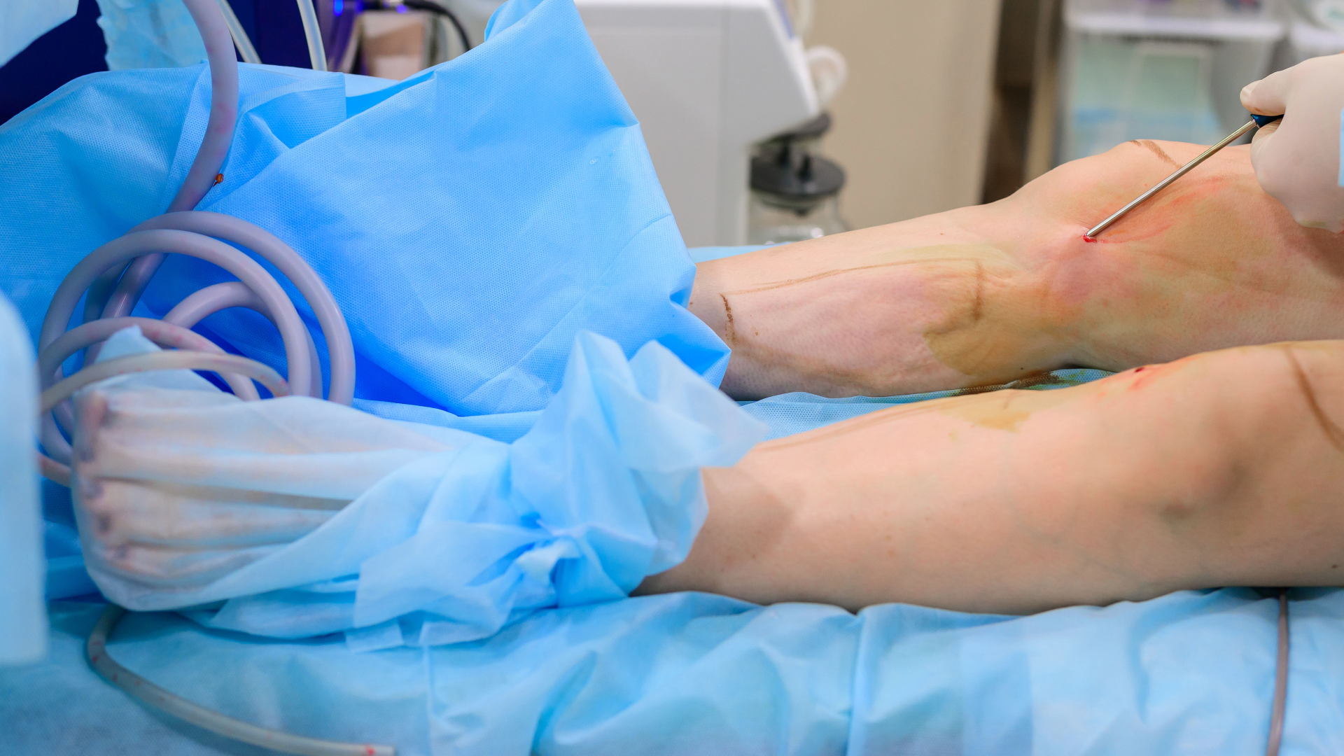
Seroma drainage after lipo is the removal of fluid that can collect under the skin after liposuction. It minimizes swelling and decreases infection risk. Drainage can be facilitated through needle aspiration, drains, and even compression garments that promote fluid movement from tissue. Timing, technique, and follow-up vary by case and surgeon. Patients typically require brief clinic visits and simple wound checks to promote healing and prevent reoccurrence.
Understanding Seromas
Seromas are pockets of clear, straw-colored fluid that accumulate under the skin after liposuction or any other procedure that extracts or disrupts a substantial amount of tissue. They occur when lymphatic channels and capillary-sized blood vessels in the treated region release lymph and serum into the void created by extracted fat or compromised tissue. Most seromas are small and carry little risk, but they can result in swelling, a soft fluctuating bulge, and discomfort at the surgery site.
Formation is common after liposuction and abdominoplasty since these procedures involve separation of large tissue planes and interruption of lymphatic drainage. Symptoms usually present themselves roughly 7 to 10 days post-surgery, but can vary. The fluid can move with motion, and some seromas will weep clear fluid through the incision or drain sites. In most cases, the body reabsorbs the fluid over weeks to months. The majority go away within a month, but some can last several months or, rarely, up to a year.
Aggressive lipo tips up the risk for seromas. When surgeons employ high-volume suction, wide undermining or power-assisted devices that disrupt more tissue, there is a bigger dead space and more lymphatic damage. That larger raw surface calls fluid to it. Consider prolonged, HD body contouring or combination cases where liposuction is combined with large-scale skin tightening. Surgeons can mitigate risk by minimizing unnecessary dissection, employing meticulous technique and placing surgical drains when appropriate.
Seromas are different from other post-op collections. A hematoma is a blood collection that is generally darker, can clot, and causes firmer swelling and more pain. An abscess indicates infection with pus, redness, spreading pain, and sometimes fever. Seromas, on the other hand, are typically painless or slightly uncomfortable and drain clear, pale fluid. If the fluid becomes cloudy or foul or the site grows tender and red, infection should be suspected and worked up.
Risk factors increase the likelihood of seroma: larger or more extensive surgery, prior chemotherapy, interrupted or poor healing, age over 65, and obesity. A correctly fitting compression garment post-op reduces the dead space and assists with lymphatic return, which decreases seroma risk. When symptomatic, enlarging and persistent, needle aspiration in clinic under sterile conditions can evacuate fluid and hasten recovery. For persistent cases, repeated aspirations, drain placement, or sclerotherapy may be required. Infected seromas need antibiotics and possible drainage.
Critical Indicators
A seroma is a pocket of clear wound fluid that sometimes develops following liposuction or abdominoplasty. Early recognition prevents delayed healing and minimizes the risk of infection or ongoing drainage. Here are the key symptoms to monitor, what they mean, and how to record them for medical consultation.
- New or worsening swelling in or around the surgical site.
- Painless, squishy mass beneath the skin that could move with motion.
- Localized discomfort, aching, or a sense of fullness
- Redness or warmth over it, particularly if pain intensifies.
- Increasing size of the swelling over the course of the day or following activity.
- Rapid buildup behind drains post-removal typically occurs 7 to 14 days post-op.
Visible Swelling
Noticeable inflammation is the most apparent symptom that fluid has accumulated under the skin. Swelling can be diffuse in the treated area or appear as a single lump. Multiple small bumps are common. Measure size and shape by taking photos from the same angle every day to compare. Recurrent swelling that doesn’t resolve after a few weeks, generally more than six to eight weeks, is called a persistent seroma and typically requires provider treatment.
Skin Fluctuation
Skin fluctuation means that if you push carefully on the swollen area, there is a wave-like motion, which indicates a fluid pocket underneath. Perform the “wave test” by tapping one side of the lump and observing for a ripple across the surface. If positive, this reinforces a diagnosis of seroma. Gentle palpation may uncover a soft, mobile pocket that moves separately from denser tissue. Document location, diameter, and if the fluctuation shifts with position. This assists clinicians in determining whether observation or aspiration is appropriate.
Localized Discomfort
Fluid-filled pockets push on nearby tissue and nerves, causing localized tenderness or aching. Pain is usually aggravated by movement, tight clothing, or direct pressure and can often be a peaty fullness instead of stabbing pain. If pain increases, the area becomes hot or shows spreading redness, suspect infection or another complication and get prompt evaluation. Small seromas may be mildly uncomfortable and can subside as the body reabsorbs the fluid over weeks, particularly with the aid of a compression garment.
Other risk factors included older age, large or repeated procedures, preoperative chemotherapy, interrupted healing and higher body mass. Clinical evaluation is necessary to exclude other etiologies and to direct management.
Clinical Assessment
A targeted clinical evaluation identifies the presence of a seroma, determines urgency, and directs subsequent steps. Examination includes inspection, palpation, basic bedside tests, imaging, and chart review. Track timing: most seromas appear within the first few weeks, commonly when drains stop, about 7 to 14 days post-op. Flatulent collections beyond 6 to 8 weeks require active treatment, not observation.
Diagnostic Ultrasound
Ultrasound is the main imaging to see fluid, to map pockets and to calculate volume. It reveals anechoic or hypoechoic collections with fine septations in several instances and aids in discriminating free fluid from hematoma or organized clot. Use ultrasound to plan aspiration. Identify the largest dependent pocket and safe needle path, avoiding vessels and the plane of recent incisions.
Correlate US findings directly with the physical exam. Soft fluctuant swelling exhibiting a discrete pocket of clear fluid on scanning supports a seroma. A complex echoic mass is more suggestive of blood or infection. Take longitudinal measurements—length, width, and depth—in millimetres and estimate volume as well for follow-up. Repeat scans at intervals to follow resolution or refilling post-aspiration and guide additional drain placement if necessary.
Ultrasound guides drainage catheter placement when serial aspirations miss. Image guidance minimizes missed pockets and diminishes the risk of damage to adjacent tissues. Document imaging reports and capture images or clips in the patient record for consistency.
Fluid Evaluation
Evaluate aspirate for color, clarity, and cell content to distinguish serous fluid from pus or blood. Serous fluid is typically straw-colored, clear to slightly hazy, and scant in cellular debris. Bloody or brown-tinged fluid indicates hematoma or surgical site bleeding. Frankly purulent fluid suggests infection and needs to be cultured and antibiotic planned.
Record the amount drained, character and any lab reports. Keep a running log: date, site, volume (mL), color, turbidity, specific gravity or protein if tested, and culture outcome. Small seromas can be noted as the body reabsorbs fluid over weeks with compression garment support. Larger collections may require serial aspiration and may take months to resolve.
Use a simple table to summarize typical fluid features:
| Characteristic | Serous Fluid | Pus/Infected | Blood/Hematoma |
|---|---|---|---|
| Color | Pale yellow | Yellow-green to brown | Red to brown |
| Clarity | Clear to slightly cloudy | Turbid | Often opaque |
| Infection signs | Absent | Often present (odor, warmth) | Variable |
| Action | Monitor/aspirate | Culture + antibiotics | Evaluate for hemostasis |
Include patient history and operative notes when interpreting results. Technique, drain duration, and intraoperative events change pretest probability. Use a checklist: history, timing, wave test, exam, ultrasound, aspiration, fluid analysis, plan to make assessment consistent and thorough.
Drainage Approaches
Drainage is selected according to seroma size, location, rate of reaccumulation, and patient factors. Small, superficial collections may be amenable to conservative management or needle aspiration. Deep, complex or persistent seromas require imaging and gradual progression to catheter drainage or surgical evacuation. Our surgeons minimize risk during the initial operation with progressive-tension (quilting) sutures, precise plane control and judicious use of surgical drains. Some even combine sutures and drains to reduce seroma formation. Keep tabs as a seroma that refills beyond six to eight weeks tends to be stubborn and should be aggressively treated rather than watched.
1. Fine-Needle Aspiration
Fine-needle aspiration employs sterile syringes and small caliber needles to extract serous fluid from small seromas. It’s rapid and can be performed in clinic, with minimal pain and no anesthesia beyond local numbing if required. Repeat aspirations are frequent when fluid reaccumulates, and repeated taps are sensible for low-volume, slow-recur seromas as the body reabsorbs fluid. Observe for reduction of swelling and symptom relief following each tap. If the pocket refills rapidly or often, escalate to catheter placement or image-guided drainage.
2. Ultrasound Guidance
Employ real-time ultrasound to detect and direct treatment at deep or irregular pockets that cannot otherwise be palpated. Imaging increases precision for aspiration or catheter placement and decreases the risk of puncturing vessels, nerves, or organs. Ultrasound further aids in volume quantification and mapping of multifocal pockets, which guides whether a single drain will be adequate or if multiple sites require intervention. Be sure to capture ultrasound findings and images in procedure notes for quality checks and future planning.
3. Catheter Placement
When aspiration fails, a small drain or catheter can be left in the cavity for continued drainage. Attach the catheter with micropore or suture and connect it to a vacuum canister or closed-suction system for continuous drainage. Monitor output daily, document fluid color and volume, and pull the catheter when drainage reduces and the pocket has collapsed. Many doctors will pull the catheter when output is minimal for 24 to 48 hours. For persistent, high-volume, or recurrent seromas, a drain can be exchanged or a minor operation to close the space can be contemplated.
4. Surgical Evacuation
Reserve surgical evacuation for sizable, chronic, infected or capsule-forming seromas that defy conservative management. The surgeon removes any fibrous capsule and closes the potential dead space, often with quilting sutures and, if deemed necessary, a new drain. A careful method reduces additional injury and decreases the chance of return. Give explicit post-op orders on wound care, compression garment usage, and infection symptoms to guide healing.
Unique Perspectives
Seroma following liposuction remains common with rates reported from as low as approximately 1% to as high as 38% depending on technique, patient factors, and how meticulously small pockets are enumerated. Surgeons emphasize three broad aims: limit dead space, reduce inflammation, and promote tissue apposition. Other polished technical maneuvers are cautious layered liposuction, smaller cannulae in certain planes, and scrupulous hemostasis to reduce bleeding that would otherwise occupy space for fluid. A few teams supplement quilting sutures or progressive tension stitches to secure flaps and seal potential spaces. These efforts reduce quantifiable seroma rates in groups, although specific decreases differ by research and patient composition.

Surgeons use simple bedside checks to judge fluid behavior. The “wave test”—a gentle tap on one side of a lump that produces a visible wave across the surface—helps distinguish fluid collections from solid nodules. A brand-new or rapidly growing swelling several weeks or months after surgery needs assessment, since long-standing seromas can wall off and become harder to treat. Imaging, usually ultrasound, often shows early collections. For example, after laparoscopic repair, 95 to 100 percent may show early seroma on imaging, yet most resolve without intervention.
Patient journeys vary by treatment course. Small seromas may resolve in 2 to 6 weeks as the body reabsorbs the fluid. A nice compression garment expedites that by compressing tissue layers so fluid cannot reaccumulate. Bigger seromas sometimes require needle aspiration, and it is not uncommon that there are several aspirations over months before the area completely dries. If a seroma persists beyond 3 months, it is called chronic. Patients experience increased clinic visits, sporadic pain, and may require surgical drainage or even scarred cavities that resist needle aspiration.
Nuances may be at play in new liposuction tech impacting seroma rates. Power-assisted and ultrasound or laser-assisted devices can decrease operative time and enhance contouring. They alter thermal and mechanical impact on tissue. Certain devices might reduce seroma through more exact fat extraction. Others might increase it if they cause more tissue damage or dead space. Current research examines device parameters and technique modifications to achieve the equilibrium between optimal adipose extraction and minimal serous drainage. Studies into steroid use demonstrate that administering steroids in the wound or shortly after surgery reduces the inflammatory response that leads to seroma formation. Protocols differ and risks should be considered.
Regional practice patterns vary. Some north NJ and Morristown clinics prefer early retention sutures and routine ultrasound follow-up. Others prefer more compression and selective aspiration. These local trends are a mirror of surgeon training, patient expectations, and resource availability.
Recovery Protocol
Outline a solid recovery protocol. A transparent recovery plan minimizes risk, facilitates tracking, and provides direction on when to obtain care.
Compression Garments
Consistent compression is the key. Compression provides consistent pressure to minimize the dead space where fluid accumulates and aids your body in reabsorbing the little seromas within 2 to 6 weeks. When selecting your recovery garments, opt for sizes corresponding to the measurements provided by your surgical team. Ill-fitting pieces can chafe, irritate skin, or simply not manage swelling. Wear garments as prescribed, typically day and night for the initial weeks, then as tolerated and healed.
Check fit every day. Check for folds, fresh redness or pain that could indicate too-tight compression or movement. If drains were placed and removed after a few days or a week, anticipate the garment becoming more crucial. Drain removal is a notorious point at which seromas form. If skin laxity or stubborn pockets persist, talk with your surgeon about tweaking sooner, not later.
Steroid injections or short courses of systemic steroids may be considered by the surgical team to suppress the inflammatory response that drives seroma formation. This is a medical risk-based decision and should be adhered to as directed by your clinician.
Activity Limits
Avoid straining and heavy lifting in the early recovery period. The recovery protocol restricting physical activity for a few weeks after lipo or tummy tuck prevents recurrence and reduces strain on repair sites. Start light walking as soon as you feel comfortable. Walking helps circulation and reduces blood clot and fluid risks without putting strain on the surgical site.
Build back up as swelling goes down and healing becomes more stable. Recovery Protocol If seroma occurs around 7 to 14 days post-op, back off to gentler movement and contact your clinic to be evaluated. Any seroma beyond 6 to 8 weeks is considered persistent and requires active intervention to prevent complications, including implant rejection, which has loss rates of 2 to 8 percent with implants.
Checklist:
- Week 0–1: Rest, drains in place if used, wear compression continually, and short walks only.
- Week 2–4: Remove drains if not already, continue compression, light walking, and avoid lifting more than 5 to 10 kg.
- Weeks 4–6: Increase low-impact exercise, monitor for recurrent swelling, and follow up with the surgeon.
- After 6 to 8 weeks, return to higher-intensity activity only after clinical clearance.
Watch for wound cleanliness and indicators of infection. Any escalating pain, redness, fever, or changing discharge merits immediate clinical attention to exclude infection or other concerns. A clinician should examine postoperative fluid to establish that a seroma exists and prepare to aspirate, drain, or intervene further if necessary.
Conclusion
Seroma after lipo can impede healing and increase the risk of infection. Early detection and quick treatment reduce those hazards. Small, clear seromas frequently drain themselves with rest, compression and follow-up visits. Larger or painful collections require sterile needle drainage and follow-up monitoring. Ultrasound aids in drainage and confirms empty pockets. Stay on top of dressing changes, wear tight compression for as long as your surgeon recommends and monitor for fever, worsening pain or cloudy drainage. An infected seroma requires antibiotics and careful attention. With prompt drainage and easy home care, the vast majority of patients regain comfort and contour within a matter of weeks. If you experience any new swelling, increasing pain, fever or strange drainage, get in touch with your provider immediately.
Frequently Asked Questions
What is a seroma after liposuction and how common is it?
Seroma drainage after lipo It’s surprisingly common and typically presents within days to weeks. In the majority of cases, the seromas are small and will resolve with appropriate care.
How do I know if I have a seroma or normal swelling?
A seroma presents as a soft, localized pocket of fluid that can fluctuate. Typical swelling tends to be more diffuse and hard. If you see a discrete, movable lump or an increasing size, inform your surgeon.
When should a seroma be drained?
Drainage should be considered for large, painful, infected, or persistent seromas that impede healing. Your surgeon decides based on size, symptoms, and the risk of complications.
What drainage methods are used for seromas?
Typical interventions are needle aspiration, drain placement, or surgical evacuation. The choice depends on seroma size, recurrence, and risk of infection. Your surgeon will describe the best option.
Is drainage painful and what anaesthesia is used?
Needle aspiration is typically rapid and only mildly painful, with local anesthesia usually sufficing. Surgical drainage may need sedation or general anesthesia. Pain is managed with medication.
What are the risks of draining a seroma?
Complications include infection, hemorrhage, scarring, and recurrence. Good technique and aftercare minimize these dangers. Discuss possible complications with your care team.
How can I prevent seroma recurrence after drainage?
Wear your compression garments as directed, avoid strenuous activity, attend follow-up appointments, and report symptoms early. Seroma drainage after lipo.
 Directions
 Call Us
 Email Us